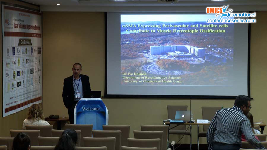
Ivo Kalajzic
University of Connecticut Health Center
USA
Title: αSMA expressing perivascular and satellite cells contribute to muscle heterotopic ossification
Biography
Biography: Ivo Kalajzic
Abstract
Heterotopic ossification is formation of bone in atypical locations including muscle. Heterotopic ossification generally occurs as a result of aberrant BMP signaling. However, further studies are required to identify BMP-responsive cells in the muscle and understand how they contribute to heterotopic ossification. Alpha smooth muscle actin (αSMA) is a marker of perivascular cells and mesenchymal progenitors that contribute to bone growth and fracture healing. We aimed to evaluate whether αSMA+ cells can contribute to bone formation during heterotopic ossification. To identify and trace cells we used αSMA promoter-driven inducible Cre (αSMACreERT2) combined with a Cre-activated tdTomato reporter Ai9 to generate SMA9 mice. Mature osteoblast/osteocytes were labeled with the Col2.3GFP reporter. To label muscle satellite cells we utilized Pax7CreERT2/Ai9 mice. Pax7Cre/Ai9 labels cells below the basal lamina consistent with a satellite cell phenotype, while SMA9 labels similarly localized cells, in addition to perivascular cells. Pax7Cre/Ai9+ satellite cells are CD45/CD31-, Sca1- but SM/C2.6+. The SMA9+ cells are CD45/CD31- and ~60% express satellite cell marker SM/C2.6. 5% express MSC markers Sca1 and PDGFRα, and a larger proportion express PDGFRβ. During BMP2-induced heterotopic ossification, SMA9+ cells comprised 28% chondrocytes and 44% of Col2.3GFP+ osteoblasts, while Pax7Cre/Ai9+ satellite cells labeled only muscle fibers. Culture of sorted cells indicated that SMA9+ and Pax7Cre/Ai9+ cells differentiated into myotubes while the negative cells do not. SMA9+ cultured cells showed the highest upregulation of osteogenic gene expression after culture in the presence of BMP2. To clarify which subset of SMA9 cells possess osteogenic potential, we subdivided SMA9+ cells based on expression of SM/C2.6 and evaluated their ability to differentiate into osteoblasts after transplantation into muscle with BMP2. We observed osteogenic differentiation of SMA9/SM/C2.6+ cells suggesting that once the satellite cell population is removed from its niche they can exhibit osteogenic potential. In conclusion, αSMACreERT2 labels a population of mesenchymal progenitor cells in the muscle that contribute to heterotopic ossification induced by BMP2. Our data indicate that the perivascular fraction of cells is responsible for heterotopic ossification, however, muscle satellite cells are capable of osteogenic differentiation after removal from their niche. Further study of these cells may indicate mechanisms by which pathogenic heterotopic ossification occurs.

